Sheffield teacher’s jaw reconstructed successfully using her fibula after rare tumour removed
24 June 2020 (Last updated: 26 Jun 2020 11:11)
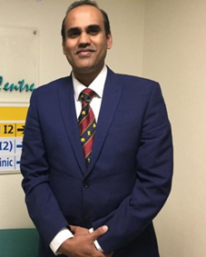
A woman with a rare tumour on her jaw has had the mass removed and her jaw reconstructed with her fibula by surgical teams led by consultant maxillofacial surgeon (OMFS) Muzzammil Nusrath from Sheffield Teaching Hospitals NHS Trust.
Muzzammil Nusrath led the oral and maxillofacial team specialists in all aspects of head and neck, facial reconstructive surgery and dental work. The operation took 13 hours to remove the non-cancerous ameloblastoma and reconstruct Gillian Wood’s jaw with a fibula free flap jaw resection. The local primary school teacher is recovering well four months after the operation, and has praised the skill of the Sheffield surgeons.
Sheffield Teaching Hospitals is a tertiary referral centre for head and neck cancer, which treats more than 200 oral and facial cancers a year.
Muzzammil Nusrath explained the surgical challenges he faced: “Gill was diagnosed with a fairly large ameloblastoma, which is a benign but aggressive tumour, that had already invaded the soft tissues of the floor of her mouth and the back of the tongue area. I needed to remove the entire affected lower jaw together with a 1cm margin of healthy bone and soft tissue to minimise the risk of the tumour coming back.”
He said that he found the cuts on the top end of the lower jaw were very close to the jaw joint and were creeping up to the skull base on the inside. So, he had to get at least a couple of screws into the area to fix the plate correctly, which had been contoured using 3D technology to minimise facial distortion.
“I needed to get the soft tissue contours of the face and the jaw line to exactly where they were before surgery, and ensure jaw function was maintained together with the bite and facial movements.”
The fibula and its blood supply can be removed safely for transplant. The leg bone blood vessels are ‘plumbed into’ the vessels of the neck with microvascular reconstruction. The muscle and soft tissue are used to build the inside of the floor of mouth and back of the tongue/cheek area.
“I am so glad that all aspects of the surgery went well and Gill has made such a good outcome and wonderful recovery. In the near future I plan to carry out her dental rehabilitation by inserting one to two implants into the new jaw.”
Gill Wood spoke about the impact of the operation’s success on her life: “Colleagues who have known me for 10 years say you can't tell I have had such a big surgery. I also met a couple of new teachers who haven't seen me before who were amazed that I look 'normal' as they have only heard about it all through my colleagues. My speech sounds completely normal, but I do have to work a little bit on not lisping - it can slip a little when I'm tired or have talked a lot! No one can believe my recovery has gone so well in such a short space of time.”
For OMFS like Muzzammil Nusrath it is vital to rebuild a patient’s aesthetics and function to retain that person’s sense of self and identity, and ensure their future quality of life: “The satisfaction of treating patients cannot be replaced by any amount of money,” he concluded.


For further information and interviews contact: Siân Evans on 020 8674 8921 / 07752 414 433 or BAOMS
Notes to editors
The British Association of Oral and Maxillofacial Surgeons (BAOMS) promotes the advancement of education, research and the development of oral and maxillofacial Surgery in Great Britain, and encourages and assists postgraduate education, study and research.
Photographs show Gill Wood before the tumour and after the tumour removal; the scar on her leg. Other photos are available.
View Other News